Dr. Bill Shear has been making regular visits to the Biology Department at East Carolina University to use their scanning electron microscope. The scope is overseen by Dr. Jason Bond, with whom Dr. Shear shares an NSF grant to study the systematic biology of millipedes. Because the SEM uses a beam of electrons instead of light, it can magnify images many, many times more than a light microscope, which is limited by the wavelength of visible light. Processors in the SEM transduce the reflected electron beams into visible light displayed on a computer monitor, and the images can be saved electronically. The enormous magnification possible is revealing bizarre surface structures in arthropods, the significance of which remains unclear. Are these features adaptive, or are they simply the structural consequences of the process by which the cuticle is constructed?
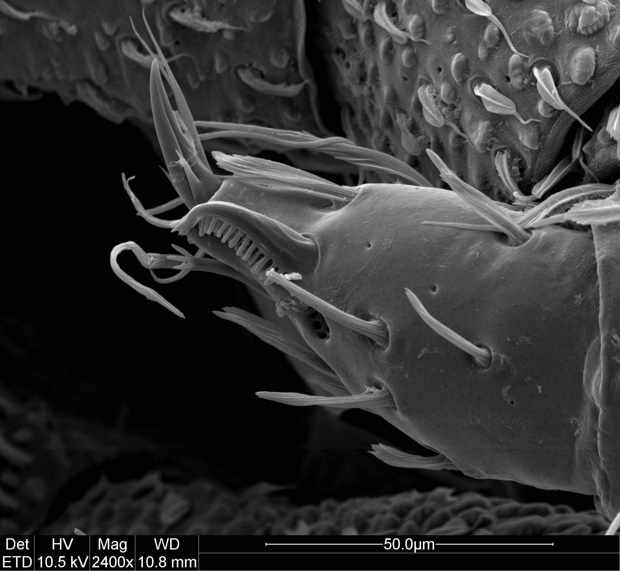
In the picture below, we are looking at the tarsus (tip segment) of the third leg of a male millipede, belonging to an undescribed genus and species. All of the spiny structures are sense organs, as is the pit with perforations in its floor. How many different ones can you find? We can hypothesize that each of them gathers unique information. Since these sensors do not occur in the females, it seems likely they have something to do with the male’s sex life, but exactly what may never be known. The whole animal is only about 7 mm long.