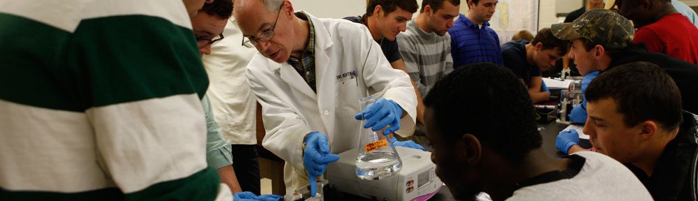
As part of the culmination of the semester-long study by H-SC Molecular Biology students into the diversity and genetics of bacteriophages, the class recently took a field trip to the University of Mary Washington to utilize their electron microscope facility. Mary Washington, like H-SC, is a member of the Howard Hughes Medical Institute’s Science Education Alliance, and UMW’s Dr. Kathy Loesser and Dr. Lynn Lewis generously shared their resources with the H-SC Molecular Biology class.
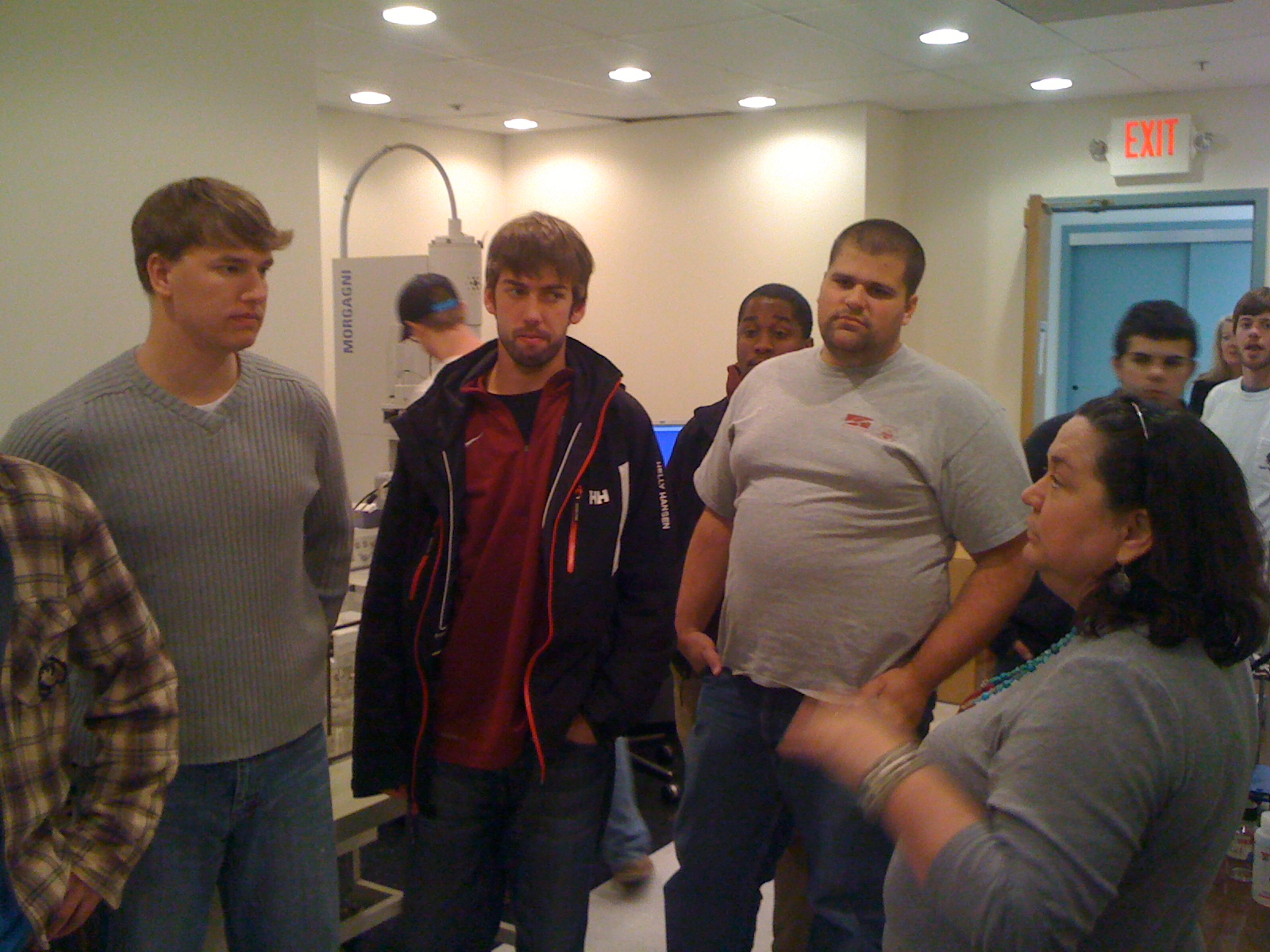
UMW's Dr. Loesser gives the class a tour of the electron microscope facility
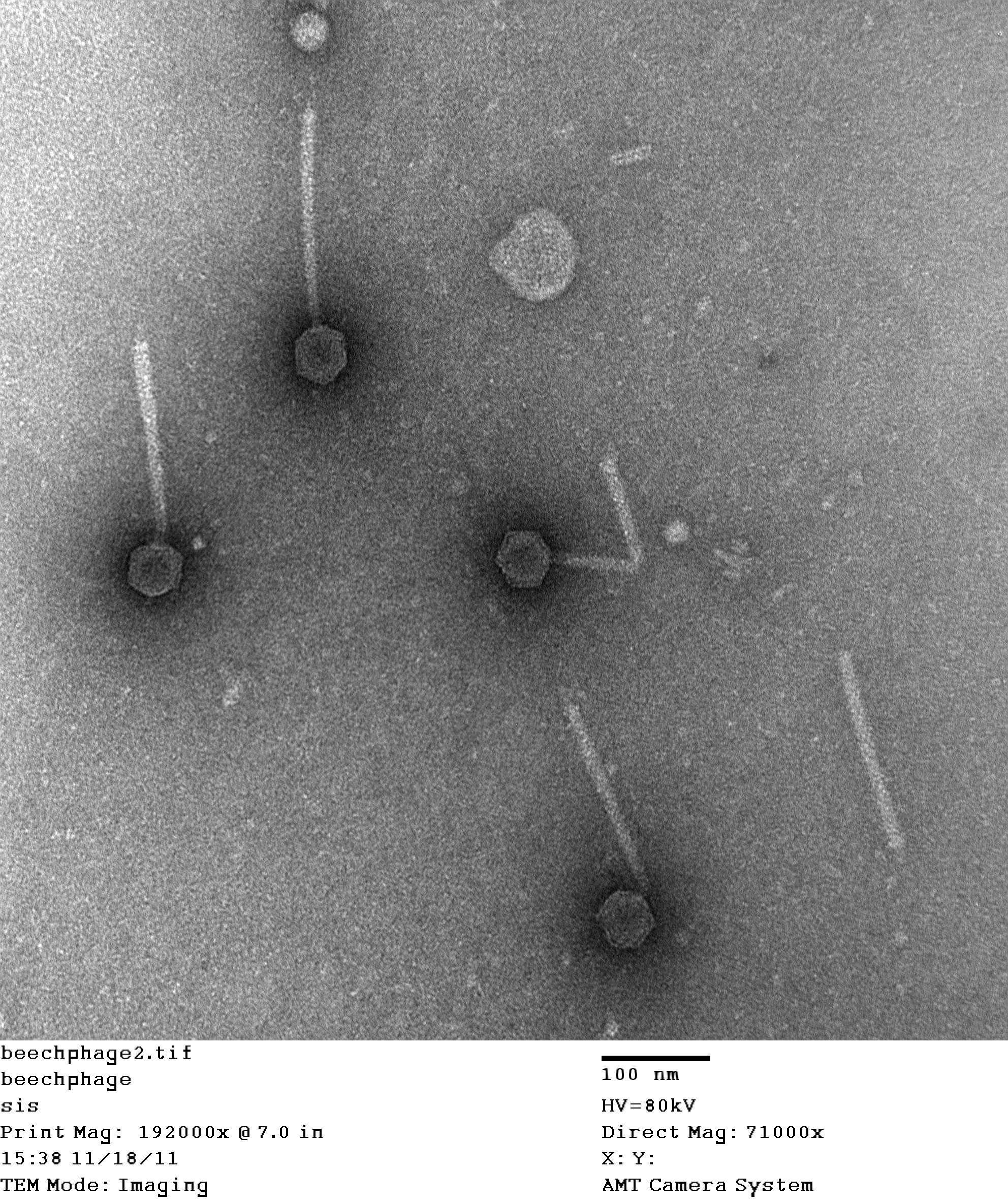
Electron microscopy can provide over 100,000-fold magnification, allowing for visualization of the viral participles that students isolated and purified over the course of the semester.
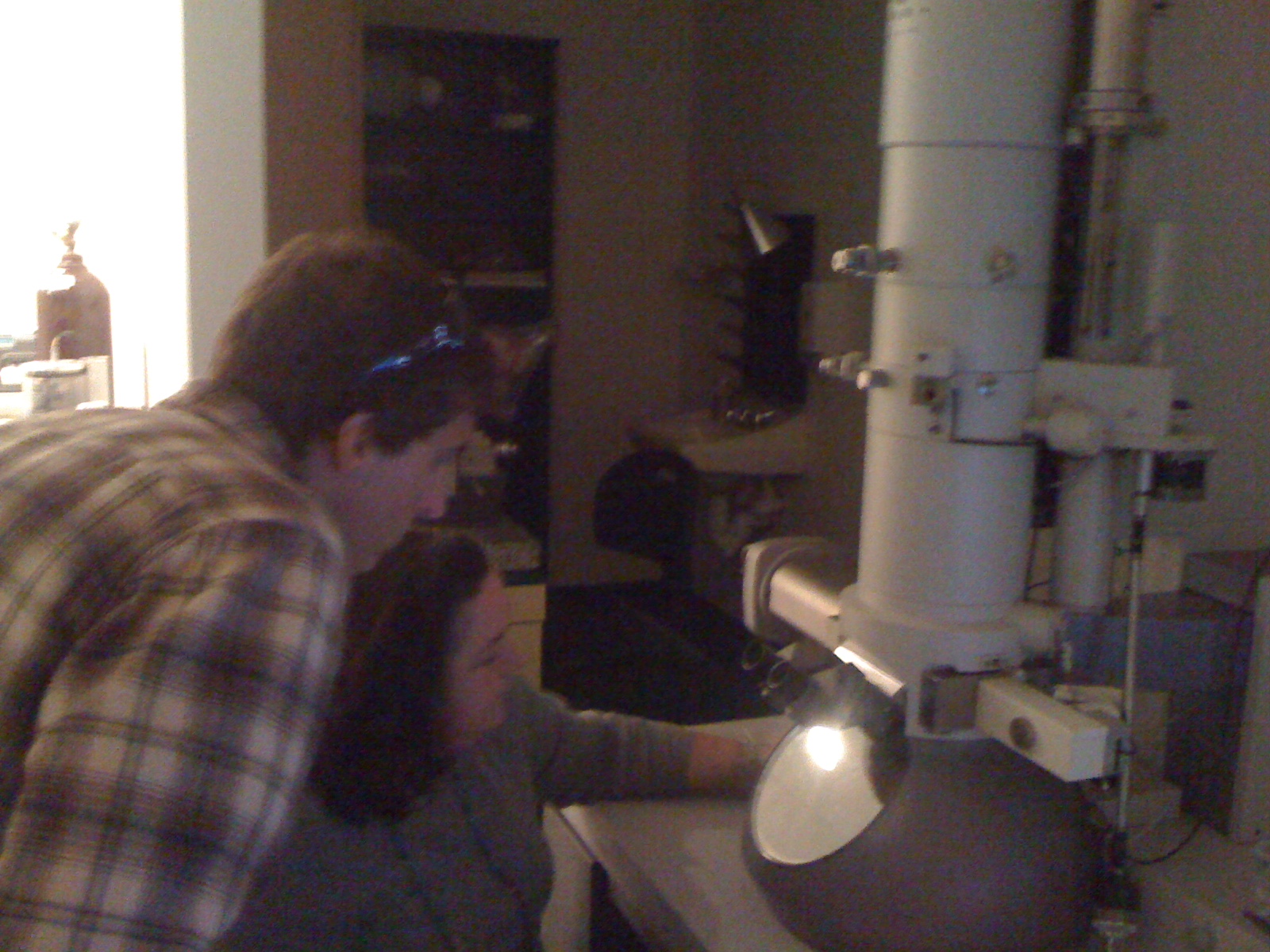
Carter Mavromatis '12 and Dr. Loesser observe "Fhageblaster", a phage isolated by Carter from Virginia Beach, VA
The electron microscope pictures provide valuable clues about the evolution of each individual phage and how they relate to phages isolated around the world by scientists at dozens of other colleges and universities.
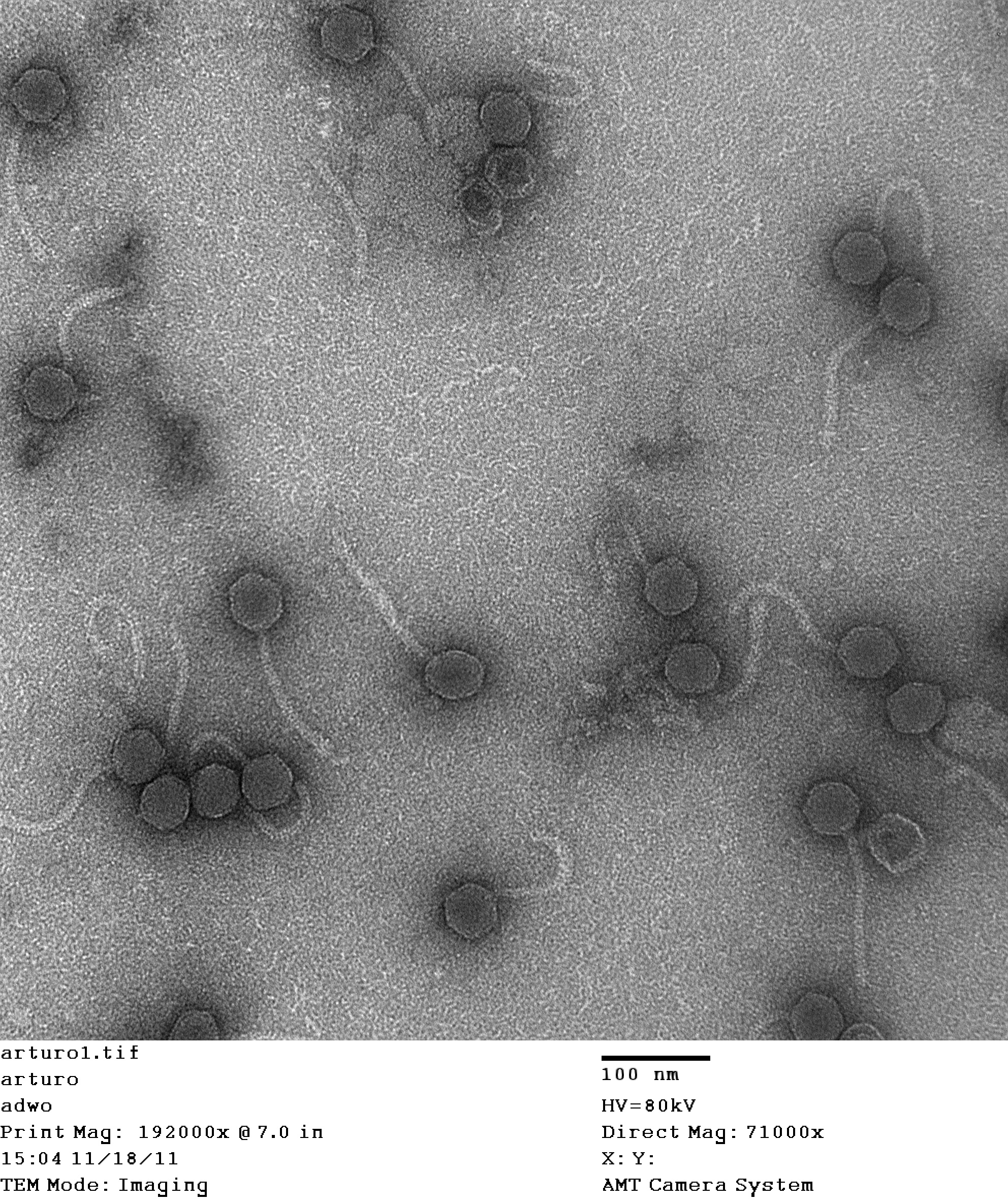
"Arturo", isolated by Duncan Oliphant '12 near the Sagebrook apartments just off the H-SC campus
"VenaBlue", isolated by Sam Smith '13 near Venable Hall on the H-SC campus