Welcome Dr. Kristin Fischer to the H-SC Biology Department

Dr. Kristin M. Fischer is very excited to return to her home state of Virginia and be the newest member of the H-SC Biology department. She earned her B.S in Biology at Virginia Tech and her interest in the medical field led her to pursue her graduate degrees from the Virginia Tech-Wake Forest School of Biomedical Engineering and Sciences. Dr. Fischer focused on tissue engineering with the goal to replace or repair damaged tissue in the body by creating a scaffold structure for cells to grow on, culturing cells on the scaffold, and implanting a functional, new tissue into a patient.

KFischer

Her graduate work at Virginia Tech and post-doctoral work at Rutgers University focused on creating a scaffold for skeletal muscle cells to grow on and culturing the skeletal muscle cells on it. The image below shows a scanning electron image of the polymeric scaffold on top and skeletal muscle cells fluorescently stained grown on it below. She completed a second postdoctoral position focusing on cardiac muscle tissue engineering at the Massachusetts Institute of Technology. In addition to her research, Dr. Fischer has previously taught a variety of courses including physiology, tissue engineering, anatomy & physiology, and introductory biology. She is looking forward to teaching in the upcoming school year.

Bio blog -2 (4)

Journal of Biomedical Materials Research Part A, 2011. 99A(3): p. 493-499.


A summer of molecular developmental neurobiology by Tyler Reekes ’17

This summer I am working with Dr. Clabough to identify a number of proteins that are vital to brain and neuronal development and may be affected by ethanol exposure. We are using a Fetal Alcohol Spectrum Disorders (FASD) mouse model and have chosen a paradigm that mimics a human third-trimester binge drinking session. Many FASD mouse models demonstrate neuronal deficits that persist into adulthood, though the specific actions of ethanol on the development of the nervous system are still not well understood, especially immediately after the ethanol exposure.

We have isolated protein from FASD and control mouse brains. We are using Western Blotting techniques to isolate specific proteins, such as Huntingtin, Brain Derived Neurotrophic Factor (BDNF), RE1-Silencing Transcription factor (REST), also known as Neuron-Restrictive Silencer Factor (NRSF), and others. We are using densitometry on ImageJ software to determine the relative concentrations of these specific proteins in important brain areas, such as the striatum and cortex.

IMG_4772


We are hoping to find changes in the expression of these genes 24 hours after ethanol exposure, and possibly a molecular correlate to a changed striatal neuronal branching that we have also observed at this early time point. The branching of these neurons is of major concern so these neurons connect to transmit signals properly. Therefore, a change in neuronal branching may translate into a similar change in behavioral, motor, or overall cognitive function that can be seen in live models.   

I have learned much about basic protein extraction and procedures by running Western Blots and interpreting results. The most important thing I have learned from this summer, is that Westerns rarely work. However, the problem solving skills I have learned from this have made me a better biologist and student in general. I cannot express my gratitude to both Dr. Clabough for all that she has taught me, but also to the Honors Council for allowing me to complete this research.

Joshua Chamberlin ’17 investigates turtle development on Hatteras Island

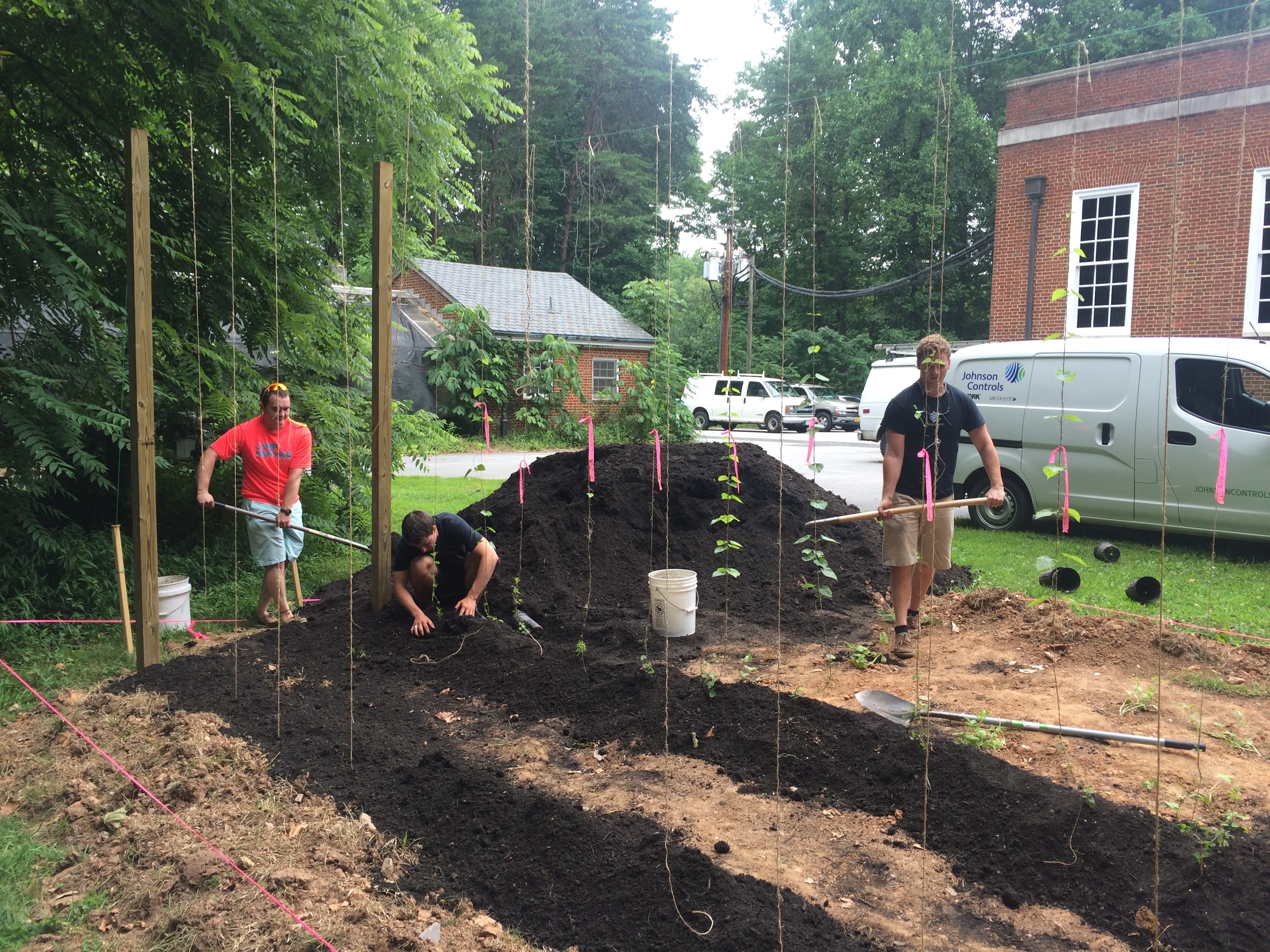
This summer I have had the privilege to join Dr. Clabough and a collaboration of people working on a project called Turtle Sense, which involves placing sensors in turtle nest to monitor when turtle hatchlings will emerge. The physical placement of an egg-shaped sensor into a turtle nest enables the relay of motion data in the nest back to a communication tower and then to a database. The motion data can then be used to predict baby turtle emergence.

IMG_5175

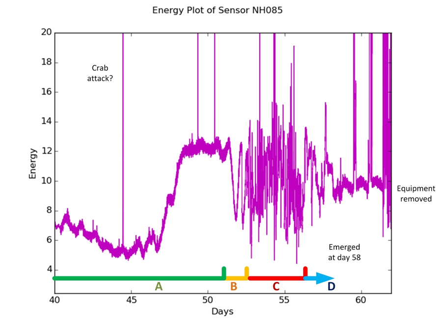
Turtle Sense data from previous years have shown that these sensors are capable of predicting when sea turtles will actually emerge from their nest within a few days. Our method of data collection can divide the motion of a sea turtle nest from day 0-60 into four phases. Phase A consist of a quiet incubation period (roughly 50 days). Phase B is a transition period that usually has some large swings with a frequency of 2-4 swings per a day. This phase ends with a quiet and low motion reading, which is often lower than anything in the preceding weeks. Phase C is the hatching activity, which is typically 4 days long and characterized by erratic, frequent motion. Phase D is a quiet period indicating emergence is imminent.

Picture1

This summer I’ve been looking at past years’ sensor data collected at Cape Hatteras to better formulate an experimentally outlined procedure for optimizing the way data is collected, as well as how the data can be used to best create a way for other people to implement the same technology in sea turtle nests in other geographic locations. We’ve seen in some data from past years that the graphs can detect non-turtle motion periods at certain points, which may indicate predation from ghost crabs, washouts from the ocean, wind manipulating the sensors, and storms.

We are working to implement a defined protocol for what to do when a nest is found, as well as specifically how each sensor should be placed into a nest to ensure less experimental error. During full nesting season, we can expect well over 200 sea turtle nests. Nest management currently requires blocking off the nest upon discovery, and after approximately 1.5 months, a larger beach enclosure is placed around the nest that essentially blocks off people from crossing that section of the beach until the nest has clearly emerged.

The best part about this research experience is that I actually get to live in Hatteras, NC for the summer. Living down here and interacting with the locals has taught me a lot about how people view sea turtles and their nesting, as well as the differing views that people have over how the beaches should be utilized regarding sea turtle nests. The nature of the project is definitely one that has proved to be a learning experience— we no longer have control over what we’re working with because we’re actually working with live animals that follow their own life cycle.

“Team Hops:” Summer study on development of optimized central Virginia hops lines

By Michael Willis ’17

“Team Hops” ( Michael Willis ’17, Traylor Nichols ’17, Gannon Griffin ’17, and Drew Elliott ’18) have been researching the hops plant specifically pertaining to downy and powdery mildews. While conducting our research we have run into several road locks where we have not had enough plants or enough growth on the plants to be able to run effective tests on the plants. This has lead us to create the Hop Garden behind Gilmer Hall. Five strains of hops are in the process of being planted in the garden: Zeus, Chinook, Cascade, Centennial, and Mount Hood. There were two sets of trellises that were erected with wire hung between them. From the wire twine was hung down for the plants to grow up on.

Willis, Nichols, and Griffin build the new hops garden and plant experimental lines.

Willis, Nichols, and Griffin build the new hops garden and plant experimental lines.

Currently several of the Chinook, and Cascade plants have climbed most of the way up the twine. After several of the plants were transferred outside we had a problem with a deer coming and eating many of the leaves off of the vines as well as biting through several vines that were climbing up the twine. The Zeus, and Chinook strains were given to us from the Virginia State University we then started to grow the plants in the greenhouse and they quickly started overtaking the light fixture in the greenhouse. Those plants were the first to be moved outside upon completing the first trellis. We then moved several plants that Ms. Jenkins had collected and rooted prior to the research from the greenhouse out into the garden. We are looking to plant more rhizomes into the new soil as soon as possible.

Dr. Laban Rutto, Virginia State University (right), visits Hampden-Sydney and provides Team Hops and Dakota Reinartz '18 (left) advice on their growth experiments.

Dr. Laban Rutto, Virginia State University (right), visits Hampden-Sydney and provides Team Hops and Dakota Reinartz ’18 (left) advice on their growth experiments.

The Hop garden will provide valuable research materials for future summer research projects as well as the Biology 151 lab. The goal is to keep the hops plants in the garden clean from infection of downy or powdery mildew.

Gene editing in zebrafish: a Hampden-Sydney student summer research experience at UAB

By Luke Bloodworth ’18

This summer I have been working with Dr. Anil Challa at the University of Alabama-Birmingham (UAB) with CRISPR, a new technology for the editing and knockout of genes.  The research we are doing at UAB with genetic engineering, given some training, would be something I would hope to be able to keep doing in Hampden-Sydney’s biology department. Dr. Challa has also expressed interest in helping us set up a zebrafish work bench so we could do our own exploration and research at HSC (He and I will present a joint seminar at H-SC in late January 2017).

The author injecting RNA samples into a polyacrylamide gel

The author injecting RNA samples into a polyacrylamide gel

Injecting fresh Zebrafish Embryos with CRISPR Cas-9 to knockout selected genes

Injecting fresh Zebrafish Embryos with CRISPR Cas-9 to knockout selected genes

So far I have used a cloud based biotech program called Benchling to locate specific spots on the amino acid coding exons of the genes we are knocking out. Taking this information, we used ensemble and CRISPR scanner to cross reference and further determine, hopefully, the proper and best binding sites for the CRISPR. Using this data, we have sequenced the proper sgRNAs (oligos) and created correlating primers for those CRISPRs. The next step will be embryo injections followed by genotyping and phenotyping.

Checking over oligonucleotides for CRISPR gene knockout in zebrafish

Checking over oligonucleotides for CRISPR gene knockout in zebrafish

I am also hoping to do a project on Argonaute (Ago). Ago is a DNA-guided DNA endonuclease, requiring no PAM sequence, and using a 24-nucleotide ssDNA with its 5′ end phosphorylated.

A summer gaining clinical experience as a certified nursing assistant

By Hayden Robinson ’18

This Summer, I have been working as a Certified Nursing Assistant. As a CNA, I have had the opportunity to be actively involved in the treatment of patients in an assisted care facility. This job not only involves basic understanding of biology, but also, an understanding of a variety of both social and scientific issues within the field of medicine. As a Hampden-Sydney Biology major, I am gaining invaluable patient contact experience, as well as a better understanding of the work necessary to monitor and treat patients in a clinical environment. With the ultimate goal of one day becoming a practicing physician, my work as a CNA allows me a very humbled and interesting perspective of the American health care system.

The author in his scrubs and ready to work

The author in his scrubs and ready to work

Summer Research Student James Ingersoll ’18 Investigates Neuronal Development

We know that ethanol exposure during development has many negative consequences for the offspring. This summer I’ve been working with Dr. Erin Clabough investigating mouse brain morphology following a one-time ethanol intoxication event in the early postnatal period for mice (which is equivalent to the third trimester of a human pregnancy). We have harvested mouse brains, followed a Golgi impregnation procedure, sectioned brains using a cryostat, followed staining procedures, used microscopes and computer programs to identify and trace medium spiny neurons, and will in the future analyze the number and types of dendritic spines.

During the Golgi staining process, we lost some usable brain sections into the wash, but in most sections, we were able to see the outline of individual neurons under the scope.

Jamie Ingersoll ’18 undertaking a Golgi staining process on mouse brain sections. The Golgi stain impregnates approximately 10% of neurons with a black pigment. We lost some usable brain sections into the wash, but in most sections, we were able to see the outline of individual neurons under the scope.

In prior study done in Dr. Clabough’s lab, we found increased branching in individual neurons in the striatum of the mouse brains immediately following a developmental ethanol exposure—which is the opposite of what we thought we’d find. We are now investigating different stages of mouse development to see when this branching phenotype disappears.

The most difficult parts of research so far have been when we were sectioning on the cryostat and it fought us for what seemed like years never wanting to yield a usable section and also the staining process where we had to watch as brain after brain slid off the slides into the unusable wash left behind.

IMG_4733

Some of our 873 brain tissue sections drying over several days prior to Golgi staining.

This experience has showed me a whole new level of patience and also how important it is to not rush the process—for example, when something like the cryostat decides it will finally cooperate, then prepare to stay for a while and crank because the next day it may not be so kind. But the best part is when everything works and we are able to see and interpret results.

We will present our research at the 2016 Society for Neuroscience meeting in San Diego in November. We hope that our research will provide insights into not just how just one binge of ethanol can affect neurons in the brain, but also how medium spiny neurons are involved in the response to ethanol and how that response may change throughout development.