Hampden-Sydney Summer Research Students Take On Melanoma

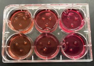
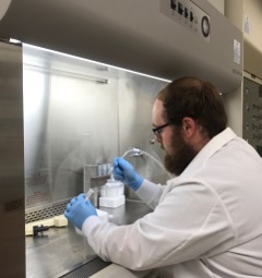
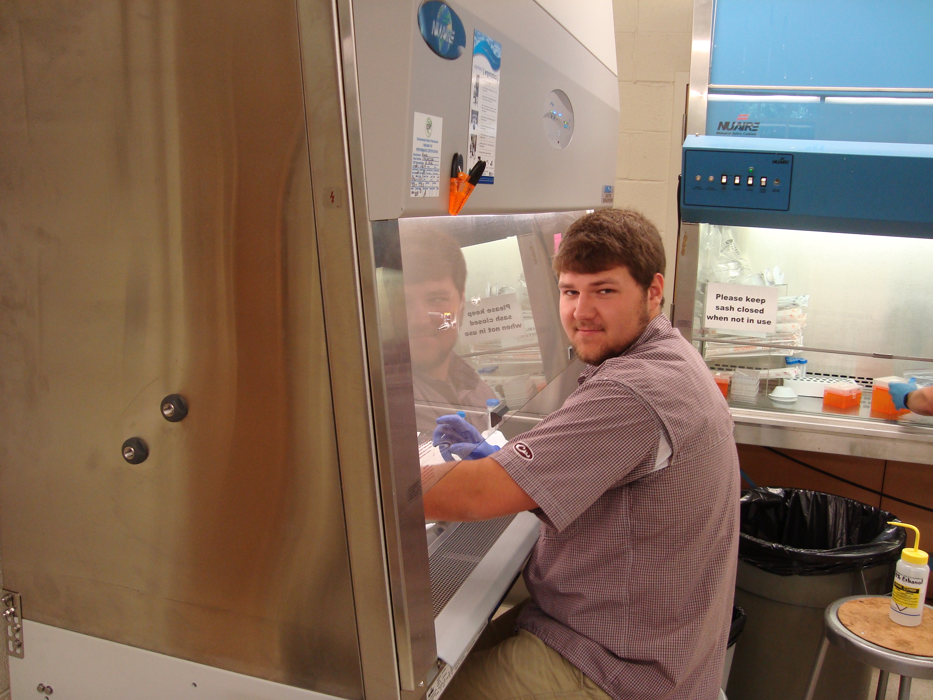
This summer, 3 rising H-SC juniors have been conducting melanoma research with Elliott Associate Professor of Biology Dr. Kristian M. Hargadon ’01.  David Bushhouse, Coleman Johnson, and Corey Williams are investigating the role of the FOXC2 transcription factor as a driver of melanoma progression.  Specifically, Corey has performed extensive phenotypic analyses of various wild-type and genetically engineered melanoma cells to study how FOXC2 expression in melanoma influences the expression of various integrins and other cell adhesion molecules on tumor cells.  In related work, Cole has focused on studying how FOXC2 expression within these melanoma cells influences tumor cell adhesion to both the extracellular matrix and lymphatic endothelial cells, processes that are instrumental in determining whether or not a tumor is able to metastasize to distant sites in the body.  In conjunction with this work, David is performing chromatin immunoprecipition studies to determine whether the regulation of cell adhesion molecule gene expression in melanoma cells that is driven by FOXC2 results from a direct interaction between this transcription factor and specific gene segments within the DNA of melanoma cells.  Collectively, this work is shaping our understanding of FOXC2’s role in melanoma progression and has the potential to identify several potential targets for cancer therapies designed to interfere with the progression of this cancer.  Cole and Corey were recently accepted into the Virginia Commonwealth University School of Medicine Early Selection Program, and David is planning to pursue a Ph.D. in the biomedical sciences.

Corey Williams '19 preparing melanoma cells for flow cytometric analysis of integrin expression.

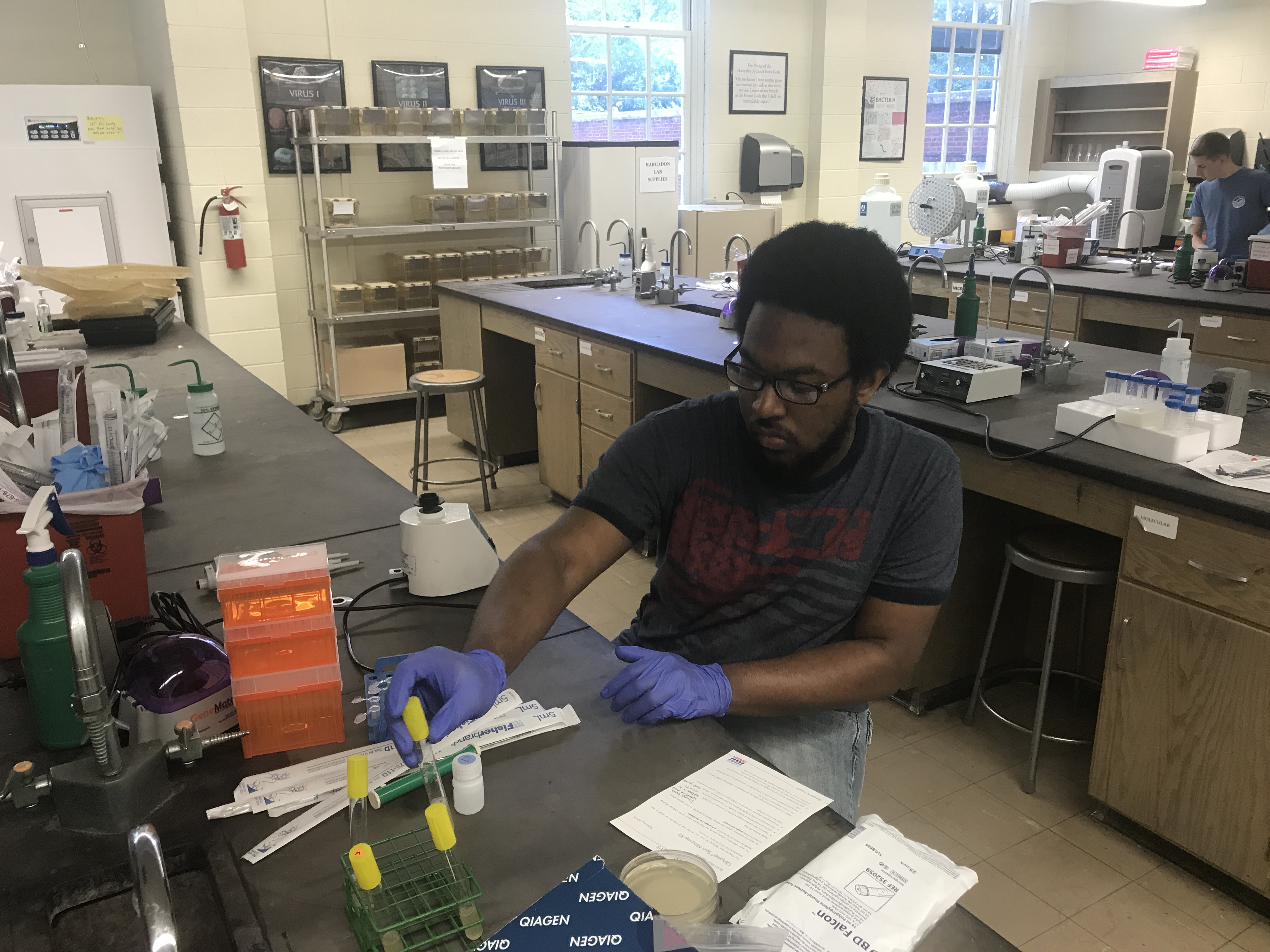
Corey Williams ’19 preparing melanoma cells for flow cytometric analysis of integrin expression!


Cole Johnson '19 setting up a tumor cell/extracellular matrix adhesion assay!

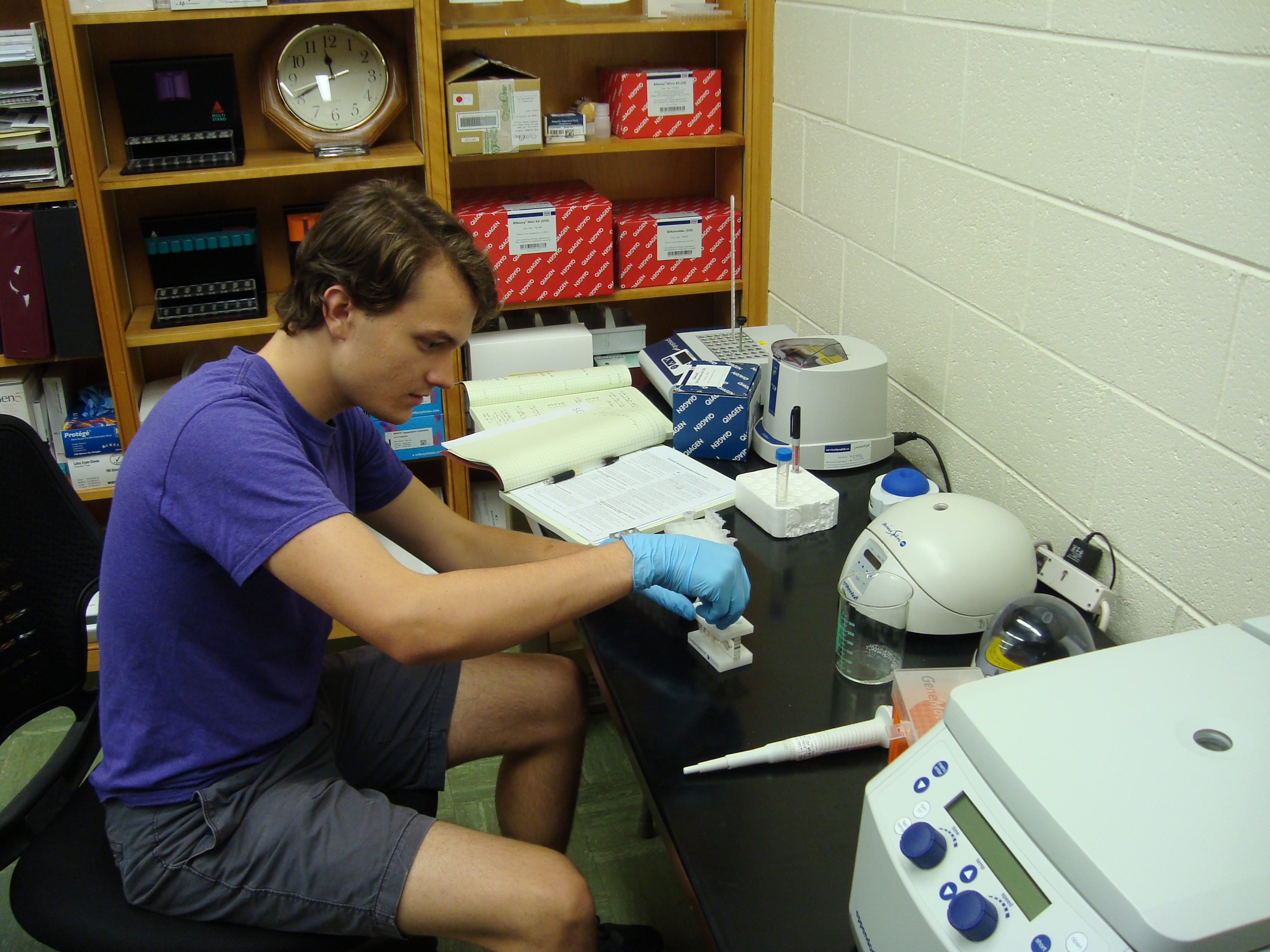
Cole Johnson ’19 setting up a tumor cell/extracellular matrix adhesion assay!


David Bushhouse '19 isolating DNA from a chromatin immunoprecipitation assay to assess target genes of the FOXC2 transcription factor!

David Bushhouse ’19 isolating chromatin from melanoma cells to assess target genes of the FOXC2 transcription factor!

Summer research in protein biochemistry

By Jason D. Pough II ’19

This summer I am working with Professor Michael Wolyniak to create an experiment for a laboratory that will be part of the future Biochemistry classes. Moreover, we are working on creating and purifying the Myf-5 Myogenic Regulatory Factor protein to assist with Professor Kristin Fischer’s research in regenerative skeletal muscle tissue as a part of creating the lab. Myf-5 is one of several Myogenic Regulator Factors that is involved with differentiating and creating muscle cells during Myogenesis. For Professor Fischer’s research, we plan to take a plasmid containing Mus musculus DNA and mutate it using site-directed mutagenesis in order to create a mutant Myf-5 protein that hopefully will aid in skeletal muscle regeneration research in conjunction with Professor Fischer’s research.

The tools of the trade for site-directed mutagenesis

The tools of the trade for site-directed mutagenesis

The overall project will aid in creating a Biochemistry laboratory by familiarizing ourselves with the techniques and methods used to carry out the Myf-5 experiment, and we will create methods and procedures for future biochemistry students to follow. One instance is with the aforementioned site-directed mutagenesis where, much like Real Time PCR, one uses primers and enzymes, yet we mutate specific sections of DNA. The mutated DNA is then inserted into a host bacterium where it will clone into a plentiful amount of bacteria with the mutated plasmid. Another instance is with the MinION Sequencer, provided by Oxford Nanopore, which uses thousands of protein pores to read the nucleotide sequence of injected DNA. The MinION Sequencer will be used to determine if the plasmids actually mutated and thus create a mutated protein. We hope that the results will not only aid Professor Fischer’s research, but also be the roots of future biochemistry laboratories and aid prospective Biology and Biochemistry and Molecular Biology majors.

Bacterial colonies after selection against ampicillin. The plasmid DNA in these cells will be sequenced to see if the desired mutation was created.

Bacterial colonies after selection against ampicillin. The plasmid DNA in these cells will be sequenced to see if the desired mutation was created.

The author prepares DNA for analysis

The author prepares DNA for analysis

H-SC summer research: developing scaffolds for mammalian tissue engineering

By Tyler McGaughey ’18

This summer I am working with Dr. Kristin Fischer to develop a porous gelatin cross-linked hydrogel scaffold for skeletal muscle tissue engineering. Through my work as an Emergency Medical Technician, I have seen numerous patients that have lost major sections of tissue. These injuries result from things like major trauma such as a car accident, violent crimes or systemic burns. These injuries have either forcefully removed the tissue or damaged it beyond repair. There are several clinical options doctors may choose: amputation, skin grafting, transplantation, or, the most interesting option, tissue engineering which is growing a new section of tissue in vitro. The ability to grow tissues outside of the body then implant them in or on humans used to be science fiction, but it is happening this summer on “The Hill”.

Dr. Fischer and I are working to answer the question of what is the best way to use the tissue engineering approach. Currently we are working with a line of mouse muscle cells called C2C12.

C2C12 cells under 400x magnification

The cells grow well given the right environment in flat sheets. However, the problem stems from layering the cells vertically. The cells in the center of the mass begin to die off due to lack of nutrients and surface area for diffusion of waste products. I intend to solve this problem by developing a gelatin scaffold for the cells to grow in. This will allow for increased diffusion and hopefully increase cell longevity.

Side view of gelatin scaffold to show thickness

Side view of gelatin scaffold to show thickness

I plan to increase diffusion to the gel by introducing pores in varying configurations. Over the last two weeks, I have tried varying number of pins per scaffold from 0-12 pins per scaffold. I have also experimented with different shapes like diagonal lines, squares, and triangles. I have concluded that the triangle formation is most likely the best formation for diffusion. I am currently attempting to print these pore inducing structures using HSC’s newest 3D printer.

3D printing a pore-inducing disk

3D printing a pore-inducing disk

The cross-linking helps the gelatin maintain its 3D structure. Cross-linking is the binding of gelatin molecules together by an enzyme called microbial transglutaminase. I have also been experimenting with different levels of microbial transglutaminase in the gelatin. More cross-linking makes the gels stiffer. There is a fine line between too much microbial transglutaminase causing the gels to rip under tension and too little microbial transglutaminase causing the gelatin to degrade too quickly.

Hydrogels with varying degrees of cross-linking in a 6-well plate

Hydrogels with varying degrees of cross-linking in a 6-well plate

In the body, muscle cells fuse together and work as one. This fusion is caused by the natural tension our muscle cells are under. In addition to introducing pores into the gel, I intend to apply a slight tension to the gels. This tension causes the muscle cells to fuse and mature in one direction as if they were in the body.

Hopefully this summer I am able to design a gelatin scaffold that helps muscle cells grow rapidly and mature.

The author working on cell culture technique in a laminar flow hood

The author working on cell culture technique in a laminar flow hood