By Tyler McGaughey ’18
This summer I am working with Dr. Kristin Fischer to develop a porous gelatin cross-linked hydrogel scaffold for skeletal muscle tissue engineering. Through my work as an Emergency Medical Technician, I have seen numerous patients that have lost major sections of tissue. These injuries result from things like major trauma such as a car accident, violent crimes or systemic burns. These injuries have either forcefully removed the tissue or damaged it beyond repair. There are several clinical options doctors may choose: amputation, skin grafting, transplantation, or, the most interesting option, tissue engineering which is growing a new section of tissue in vitro. The ability to grow tissues outside of the body then implant them in or on humans used to be science fiction, but it is happening this summer on “The Hill”.
Dr. Fischer and I are working to answer the question of what is the best way to use the tissue engineering approach. Currently we are working with a line of mouse muscle cells called C2C12.
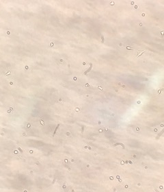
C2C12 cells under 400x magnification
The cells grow well given the right environment in flat sheets. However, the problem stems from layering the cells vertically. The cells in the center of the mass begin to die off due to lack of nutrients and surface area for diffusion of waste products. I intend to solve this problem by developing a gelatin scaffold for the cells to grow in. This will allow for increased diffusion and hopefully increase cell longevity.

Side view of gelatin scaffold to show thickness
I plan to increase diffusion to the gel by introducing pores in varying configurations. Over the last two weeks, I have tried varying number of pins per scaffold from 0-12 pins per scaffold. I have also experimented with different shapes like diagonal lines, squares, and triangles. I have concluded that the triangle formation is most likely the best formation for diffusion. I am currently attempting to print these pore inducing structures using HSC’s newest 3D printer.
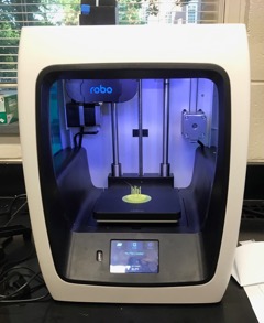
3D printing a pore-inducing disk
The cross-linking helps the gelatin maintain its 3D structure. Cross-linking is the binding of gelatin molecules together by an enzyme called microbial transglutaminase. I have also been experimenting with different levels of microbial transglutaminase in the gelatin. More cross-linking makes the gels stiffer. There is a fine line between too much microbial transglutaminase causing the gels to rip under tension and too little microbial transglutaminase causing the gelatin to degrade too quickly.
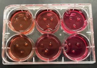
Hydrogels with varying degrees of cross-linking in a 6-well plate
In the body, muscle cells fuse together and work as one. This fusion is caused by the natural tension our muscle cells are under. In addition to introducing pores into the gel, I intend to apply a slight tension to the gels. This tension causes the muscle cells to fuse and mature in one direction as if they were in the body.
Hopefully this summer I am able to design a gelatin scaffold that helps muscle cells grow rapidly and mature.
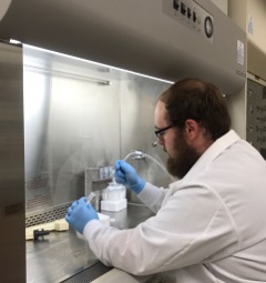
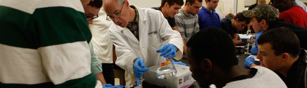
The author working on cell culture technique in a laminar flow hood